| Source: Florida Atlantic University | Released: Tue 18-Jul-2006, 09:00 ET |
Fractal Theory to Diagnose and Treat Breast Cancer
| Libraries Medical News |
Keywords BREAST CANCER, MRI, MAMMOGRAPHY, ULTRASOUND, FRACTALS, FRACTAL THEORY, DIAGNOSIS OF BREAST CANCER, TREATMENT OF BREAST CANCER | |
|
Contact Information Available for logged-in reporters only | ||
|
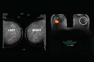
Description Fractal theory provided the basis for a unique software platform program that has been developed for use in conjunction with MRI, and is showing great promise in the early diagnosis and treatment of breast cancer. Using this unique method, a study has shown that in over 30 percent of patients there were additional tumors in the same breast, and in almost 10 percent of the patients there were tumors in the opposite breast. These tumors were not previously found using mammography or ultrasound. | ||
Newswise — Researchers from Florida Atlantic University, the Center for Breast Care at the Women’s Center at Boca Raton Community Hospital, and MeVis, The Center for Diagnostic Systems and Visualization at the University of Breman, Germany have developed new techniques to aid clinicians in the diagnosis and treatment of breast cancer. They have developed and piloted a unique software platform utilizing computational clinical imaging techniques for the analysis and display of serial-time MRI, which is showing great promise in the early detection and treatment of breast cancer.
These FDA-approved techniques were developed at MeVis, The Center for Diagnostics Systems and Visualization, under the direction of Dr. Heinz-Otto Peitgen, who is also a faculty member in the department of mathematical sciences at FAU’s Charles E. Schmidt College of Science. Peitgen used the mathematical concept of fractals to begin developing this unique software.
“Fractals are large, irregular geometric patterns made up of infinitely smaller, but identical, irregular patterns,” said Peitgen. “Fractal theory provided an appropriate platform upon which to build the software program because the ducts within human breast tissue have fractal properties.”
Breast MRI is a relatively new tool used by physicians to diagnose breast cancer as an adjunct to conventional mammography. Breast MRI displays the behavior of a cancerous lesion in three dimensions and approaches a nearly 100 percent accuracy rate in the detection of invasive cancer. In contrast, mammography provides a two-dimensional view of the breast and surrounding tissue and only detects 80 to 85 percent of tumors. One of the main strengths of MRI is its precise delineation of soft tissue and its ability to image the breast in fine sections dynamically by taking multiple MRI images over time. The percentage of medical centers doing breast MRI is small, but growing.
A recent study spearheaded by Dr. Kathy Schilling, medical director of Imaging and Intervention at the Center for Breast Care at the Women’s Center at Boca Raton Community Hospital, was published in The American Journal of Radiology and entitled “Assessment of Suspected Breast Cancer by MRI – A Prospective Clinical Trial Using a Combined Kinetic and Morphologic Analysis.” Findings of this study showed that in over 30 percent of patients there were additional tumors in the same breast, and in almost 10 percent of the patients there were tumors in the opposite breast.
“These tumors were not found using mammography or ultrasound,” said Schilling. “We also found a resulting change in the course of treatment in nearly 25 percent of patients undergoing surgery for newly diagnosed breast cancer.” In addition, findings from this study showed that MRI directed biopsies using computational clinical imaging led to definitive conclusions, demonstrating the clinical utility of this unique approach.
According to Dr. Roger Goldwyn, faculty member in FAU’s department of mathematical sciences and director of the proposed Center for the Development of Computational Clinical Imaging at FAU, “Our approach is to use innovative image-processing tools to find additional tumors and to help determine patient management outcomes. These techniques have also led to biopsies directed by these image-processing tools, surgical planning modifications, and monitoring effectiveness of chemotherapy.”
The Center for Breast Care at Boca Raton Community Hospital is a high volume center, handling more than 45,000 cases per year and finding more than 320 new breast cancers annually. In conjunction with FAU, the Center for Breast Care is developing their expertise in the use of computer-aided clinical imaging to assist clinicians nationwide. The integration of this software into general imaging departments involved in breast cancer diagnosis will enable better care, a reduction in costs for unnecessary surgeries, and ultimately result in improved patient survival. The Center for Breast Care is also working with experts at FAU in the university’s medical school program to further develop these specialized tools and continue to evaluate them in pilot studies to examine their ability to save lives.
“Our researchers at FAU will continue to conduct clinical evaluations and collaborate with the Center for Breast Care and MeVis to further develop these tools for use by clinicians in more routine settings in order to have a wider impact on patient care,” said Dr. Larry F. Lemanski, vice president for research at FAU.
American women have a one in nine chance of developing breast cancer during their lifetime. Early detection is the single most effective tool in fighting breast cancer. The sooner the cancer is detected, the more options a woman has for treatment and the better her chance for survival. Methods for detection include mammography—a safe, low-dose x-ray of the breast—that can be a vital tool in helping to discover small lumps up to two years before they can be felt in a physical exam. With mammography, however, only 80 to 85 percent of tumors are detected, leaving 15-20 percent of tumors undetected. These undetected tumors will enlarge and become more lethal before they are identified in a later screening or when they become clinically apparent.
View Video: mms://131.91.97.42/axon/EarMarked_revised.wmv
- FAU -
Florida Atlantic University opened its doors in 1964 as the fifth public university in Florida. Today, the university serves 26,000 undergraduate and graduate students on seven campuses strategically located along 150 miles of Florida's southeastern coastline. Building on its rich tradition as a teaching university, with a world-class faculty, FAU hosts eight colleges - the Dorothy F. Schmidt College of Arts & Letters, the Charles E. Schmidt College of Science, the Christine E. Lynn College of Nursing, the Harriet L. Wilkes Honors College, and the Colleges of Business, Education, Engineering & Computer Science, and Architecture, Urban & Public Affairs.
