| Courtesy of Motorola Life Sciences |
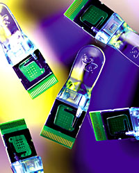 It's Bioelectric! Academic and industrial
researchers are developing a dizzying array of strategies to link
biological macromolecules and electronic circuitry. Motorola's
eSensor, for example, utilizes a ferrocene-conjugated signaling
oligonucleotide that, when captured on the chip, produces a current
whose magnitude is proportional to the quantity of probe that is
bound.
|
Researchers in the rapidly progressing field of molecular bioelectronics are coupling biomolecules with some unlikely mates from the "non-biotic" side of town: electronic transducers. Communication between these odd partners from the biologic and nonliving worlds is effected by the passage of electrons. The potential rewards for these innovations are considerable. Bioelectronic systems could one day be employed in a wide variety of apparatuses, including biosensors, devices for driving biotransformations, biofuel cells, and perhaps even biocomputers.3-5
The New Biosensors
The biomolecular and electronic components are electrically connected via molecular electron transfer relays. The specifics of how this is accomplished vary greatly, and can involve biomolecules that are inherently capable of electron transfer, such as redox enzymes or exogenous electronic mediators, which can be attached to the electrode or a biomolecule, or can act as diffusional electron couriers. Biomolecules and electronic elements are joined using any of a variety of techniques, but the redox-active portions of the biomolecules and other electroactive components must be closely and appropriately positioned to enable the electronic connection.
Today's bioelectronic sensors generally consist of an electronic transducer whose surface is engineered to support a biomolecule-bearing film, monolayer, or multilayer of controlled thickness.2-4 Often, these substrates are functionalized with nucleic acids, proteins, or ligands that recruit their binding partners through affinity interactions. The sheer diversity of bioelectronic strategies precludes a comprehensive discussion, but a few examples are presented below.
Itamar Willner, chemistry professor at Hebrew University of Jerusalem, has developed a general strategy for quantifying the substrates of cofactor-dependent redox enzymes. A relevant enzyme depleted of its cofactor—an apo-enzyme—is reconstituted on electrodes functionalized with relay moieties linked to the cofactor.3,4 In one example, Willner's group fashioned a glucose biosensor by crosslinking apo-glucose oxidase to an electrode-bound monolayer consisting of pyrrolquinoline quinone (PQQ)-flavinadenine dinucleotide (FAD). When the immobilized enzyme oxidizes glucose, electrons are shuttled from its cofactor FAD to PQQ to the electrode.
"This technology could be employed in implantable devices for continuous blood glucose monitoring," comments Willner. "We have also constructed a noncompartmentaalized biofuel cell employing electrodes functionalized with PQQ-FAD- glucose oxidase and cytochrome c-cytochrome oxidase. This type of device could be used as an implantable energy source, in which blood glucose is used to fuel the generation of electrical energy."
The group's other interests include the development of reusable optobioelectronic immunosensors, and biosensors for DNA analyses.3,4 Amplified electronic transduction of DNA sensing can be effected using biotinylated probes to recruit enzyme conjugates, which measurably decrease electron transfer by depositing insoluble precipitate on the electrode.3
Homme Hellinga, associate professor of biochemistry at Duke University Medical Center in Durham, NC, and colleagues have recently developed a versatile, protein conformation-based strategy for chemoresponsive bioelectronic systems.5,6 The ligand-induced, hinge-bending motions of bacterial periplasmic-binding proteins (bPBP) are exploited to switch an electronic current on or off.
Hellinga's group has constructed a family of four bioelectronic sensors with differing specificities: two for the sugars maltose and glucose; one for the amino acid glutamine; and one for zinc ions.6 These sensors employ different bPBPs to dictate specificity: maltose binding protein (MBP), superfamily members specific for glucose or glutamine, or a MBP-based construct engineered to respond to Zn(II). Constructs with other specificities could potentially be employed as well.
The bPBP is tethered to an electrode surface through a genetically engineered construct bearing an oligohistidine tag. A Ru(II) redox reporter group is then conjugated to the bPBP at a modified cysteine residue located near the electrode. In the absence of the target ligand, the Ru(II) reporter is proximal to the electrode, and a current is generated. Ligand binding interrupts this electrical signal by inducing a conformational change that moves the reporter group away form the electrode. The resultant decrease in current is proportional to the population of ligand-bound protein, and thus to ligand concentration.
Another nonenzymatic tactic involves the use of responsive transmembrane channels. One type of device employs gramicidin ion channels that are controlled by a pair of antibodies.7,8 A lipid membrane containing two sets of gramicidin half-channels is tethered to a gold electrode: those in the inner membrane leaflet are immobilized to the electrode, and those in the outer leaflet are mobile. The space between the electrode and the membrane contains an "ion reservoir." When a potential is applied, ions flow from the reservoir to the external solution if functional ion channels are present. Antigens prevent ion channel formation via the crosslinking of two different monospecific antibodies, one bound to the mobile gramicidin half-channels, and one bound to an immobilized membrane-spanning lipid. This method can be adapted for other applications by replacing the antibodies with relevant binding partners, such as oligonucleotide probes for DNA detection.
Hybrid DNA Biosensors
Synthetic DNA probes corresponding to mutational "hot spots" are attached to a gold electrode. After hybridization with samples containing patient DNA, methylene blue (MB+), a redox-active DNA intercalator, is added along with a ferricyanide [Fe(CN)6]3- salt. MB+ serves as an electron acceptor when current flows from the electrode. The reduced intercalator then reduces [Fe(CN)6]3- to regenerate MB+, which goes on to accept more electrons in a catalytic cycle.
Only intact duplexes formed from normal DNA allow optimal current flow. Those formed from mutated DNA will contain at least one mismatch, resulting in a readily detected abatement of current. GeneOhm has implemented this technology in a microchip-based platform. "We are excited about applying this technology to SNP diagnosis and advancing genetic analysis as a convenient and accessible tool for patient management," comments Barton.
Schaumburg, Ill.-based Motorola Inc. offers the eSensor™ DNA detection system through its Pasadena, Calif.-based Life Sciences division. This system employs two single-stranded DNA probes: one for capturing and immobilizing the target to the electrode surface, and one for bioelectronic signaling.10 The capture and signaling probes are complementary to discrete, adjacent portions of the target DNA or RNA sequence.
A gold electrode in a printed circuit board is coated with a self-assembled monolayer (SAM) containing unlabeled capture probes. Signaling probes are free in solution, and contain ferrocene-modified nucleotides that serve as electronic labels. Binding of target DNA to both probes brings the ferrocene moieties in close proximity to the electrode surface. When an appropriate voltage is applied, the hybridized signal probes are oxidized from Fe(II) to Fe(III) as they transfer electrons through the SAM to the electrode. The current generated by the oxidation of ferrocene is directly proportional to the number of ferrocene moieties immobilized at the electrode surface.
"Conventional high-density DNA microarray- or chip-based approaches often employ fluorescent probes and optical readers. Though powerful, most DNA chip methodology is expensive, and perhaps most practical for use in large-scale efforts typical of Big Pharma," observes Daniel Farkas, director of clinical diagnostics at Motorola Life Sciences. "Our biosensor-based technology provides a flexible and economical alternative for applications that require relatively fewer interrogations, such as molecular diagnostics or environmental screening," he adds.
The company recently announced two clinical collaborations. Last May, Wayne Grody, medical director of the molecular genetics laboratory at the University of California at Los Angeles, and his team began evaluating the eSensor system for use in detecting mutations associated with hemochromatosis, a hereditary disorder of iron metabolism. Sanofi-Synthélabo Inc. of Paris is using eSensors to simultaneously detect mutations in three members of the cytochrome P450 superfamily to evaluate candidate therapeutics at it's Malvern, Pa.-based North American R&D headquarters.
| Courtesy of Toshiba Corp. |
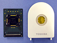 Toshiba's hepatitis C SNP typing chip
|
Tokyo-based Toshiba Corp. has developed an electrochemical DNA chip for the SNP typing of patients infected with hepatitis C. These chips will be used to identify patients most likely to respond to interferon therapy. Capture probes are immobilized onto gold electrodes through a SAM. After the hybridization reaction to the target DNA, Hoechst 33258, an electrochemically active dye that specifically binds the minor groove of double-stranded DNA, is added. When an appropriate potential is applied, the oxidative current from the dye is proportional to the amount of bound target DNA.
According to Toshiba, this system has a number of advantages, including its amenability to miniaturization and facile large-scale chip production. The company also plans to apply this technology towards the development of other individualized treatment protocols.
|
Courtesy of Xanthon Inc. |
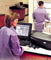 Xanthon's Xpression Analysis System
|
Xanthon Inc. of Research Triangle Park, NC, plans to release its Xanthon Xpression Analysis System™, later this year. Based on the electrochemical oxidation of the guanine base moieties of target nucleic acids,11 this system commercializes technology discovered under the direction of Holden Thorp, professor of chemistry at the University of North Carolina at Chapel Hill.
Electrodes are functionalized with synthetic oligonucleotide probes that contain the nonphysiological base inosine instead of guanine to minimize background signal. After introducing the sample and washing away nonhybridized material, a redox-active mediator of appropriate potential, usually ruthenium tris (2,2'-bipyridine), is added, and an appropriate voltage is applied. Each mediator molecule is oxidized from Ru(II) to Ru(III) as it transfers an electron to an electrode. In the absence of guanine, each mediator molecule is oxidized only once, but when a guanine-containing nucleic acid target is present, a current-enhancing catalytic cycle is created. "The catalytic cycle provides an in situ amplification of the signal and a direct readout based on guanine," explains Thorp. Neil Moore, Xanthon's vice president of business development, adds, "Although our first commercialized system is for microplate assays, the underlying technology can be readily adapted for use in other formats, such as biochips for genomic research, and even hand-held devices for bioprocess control or medical diagnostics."
BioE & Friends
The use of chemical and genetic engineering approaches to modify protein structure and function has much to offer today's biomolecular engineers as well.2,7 For example, *-hemolysin pores can be fitted with internal, noncovalently-bound molecular adapters that "program" them for a range of sensing functions.13 Useful protein modules are also being made from scratch; for example, a nitrate-sensing electrode has been assembled by crosslinking nitrate reductase and a de novo four-helix-bundle protein reconstituted with Fe(III)-protoporphyrin.4 Advances in methods for modifying surfaces with biomaterials in microscale patterns are also paving the way towards devices with expanded capabilities, such as self-calibrated biosensors and biosensor arrays.4
Though this article focused on molecular bioelectronic sensors, researchers are also combining biomolecules with optical, magnetic, gravimetric, and mechanical elements.4,6,14 These biomolecular engineers aren't the only ones forging devices from biologic and non-biologic components; other modern day pioneers are developing cell-based hybrids, such as neuroelectronic circuitry and whole-cell biosensors.15-17 As the boundaries between biological, cellular, chemical, and physical sciences diminish, expect more technological innovations, with a concomitant upsurge of new biotech companies to commercialize them.
1. R.F. Service, "Molecules get wired," Science, 294:2442-3, Dec. 21, 2001.
2. G. Gilardi, A. Fantuzzi, "Manipulating redox systems: Application to nanotechnology," Trends in Biotechnology, 19:468-76, November 2001.
3. I. Willner, B. Willner, "Biomaterials integrated with electronic elements: En route to bioelectronics," Trends in Biotechnology, 19:222-30, June 2001.
4. I. Willner, E. Katz, "Integration of layered redox proteins and conductive supports for bioelectronic applications," Angewandte Chemie, International Edition in English, 39:1180-218, 2000.
5. I. Willner, "Protein hinges for bioelectronics," Nature Biotechnology, 19:1023-4, November 2001.
6. D.E. Benson et al., "Design of bioelectronic interfaces by exploiting hinge-bending motions in proteins," Science, 292:1641-4, Aug. 31, 2001.
7. H.W. Hellinga, J.S. Marvin, "Protein engineering and the development of generic biosensors," Trends in Biotechnology, 16:183-9, 1998.
8. B.A. Cornell et al., "A biosensor that uses ion-channel switches," Nature, 387:580-3, 1997.
9. E.M. Boon et al., "Mutation detection by electrocatalysis at DNA-modified electrodes," Nature Biotechnology, 18:1096-1100, 2000.
10. R.M. Umek et al., "Electronic detection of nucleic acids: A versatile platform for molecular diagnostics," Journal of Molecular Diagnostics, 3:74-84, May 2001.
11. H.H. Thorp, "Cutting out the middleman: DNA sensors based on electrochemical oxidation," Trends in Biotechnology, 16:117-21, 1998.
12. A.M. Leone et al, "An ionic liquid form of DNA: Redox-active molten salts of nucleic acids," Journal of the American Chemical Society, 123:218-22, Jan. 17, 2001.
13. L.Q. Gu et al., "Capture of a single molecule in a nanocavity," Science, 291:636-40, January 2001.
14. J. Wang, "From DNA biosensors to gene chips," Nucleic Acids Research, 28:3011-16, 2000.
15. G. Zeck, P. Fromherz, "Noninvasive neuroelectronic interfacing with synaptically connected snail neurons immobilized on a semiconductor chip," Proceedings of the National Academy of Sciences, 98:10457-62, Aug. 28, 2001.
16. J.O. Winter et al., "Recognition molecule directed interfacing between semiconductor quantum dots and nerve cells," Advanced Materials, 13:1673-7, November 2001.
17. A. Offenhäusser, W. Knoll, "Cell-transistor hybrid systems and their potential applications," Trends in Biotechnology, 19:62-6, February 2001
The Scientist 1[6]:38, Mar. 18, 2002